Radioimmunoassay (RIA)
Radioimmunoassay (RIA) is a powerful analytical technique used for the quantification of specific molecules, including antigens, antibodies, hormones, and other analytes present in biological samples. Unlike conventional assays, RIA utilizes the unique properties of radioactive isotopes to trace and measure the concentration of target molecules. This method offers a high level of sensitivity and accuracy, making it particularly valuable in various scientific investigations and clinical diagnostics.
Principle of Radioimmunoassay (RIA)
Radioimmunoassay is based on the competitive binding of a radioactive-labelled antigen (tracer) and the unlabelled antigen in the test sample to a limited amount of antibody. The amount of detected radioactivity of the bound tracer inversely correlates with the concentration of the target antigen in the sample.
Hot antigen: Hot antigen also known as “tracer” refers to an antigen that has been radioactively labelled.
Cold antigen: Cold antigen refers to the unlabeled antigen present in the test sample that competes with the labelled (hot) antigen for binding to the limited binding sites on the antibody.
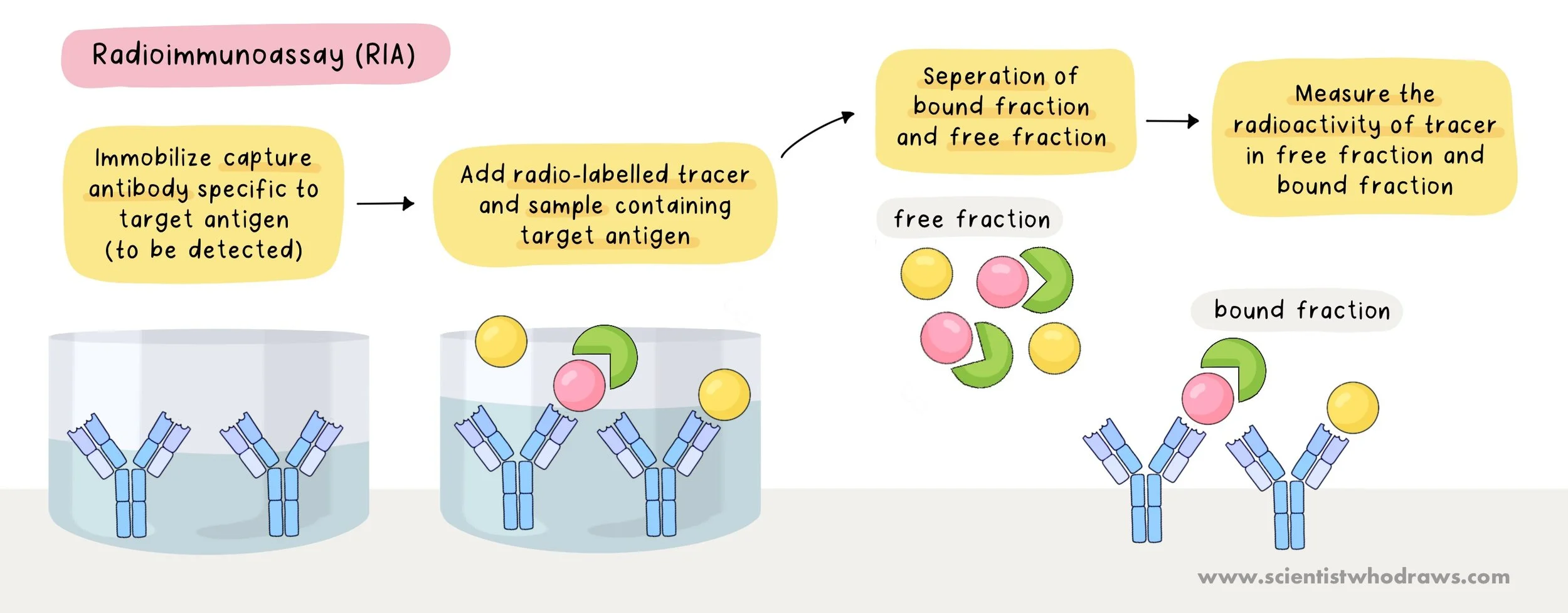
Figure: Procedure for Radioimmunoassay (RIA)
✏️ Procedure for Radioimmunoassay:
Preparation of Hot antigen or Radioactive Tracer: The antigen of interest is labelled with a radioactive isotope, such as iodine-125 (I-125), to create a radioactive tracer or ‘hot antigen’.
Coating the microplate with Antibody: The inner surface of the assay tubes is coated with an antibody specific to the target antigen. This antibody will capture both the radioactive tracer and the unlabeled sample.
Addition of Hot antigen and Cold antigen: The hot antigen or ‘tracer’ and the test sample containing cold antigen are simultaneously added to the microplate.
Incubation: Incubate the tubes to allow the competitive binding between the radioactive tracer antigens and the unlabelled samples antigens for binding sites on the antibody.
Separation of Bound and Free Fraction: The bound and free fractions are separated using a separation method like precipitation or filtration.
Radioactivity Measurement: The radioactivity of both the bound and free fractions is measured using a gamma counter. The ratio of bound to free radioactivity provides a quantitative measure of the concentration of the target antigen in the sample.
Data Analysis: The amount of detected radioactivity of the bound fraction inversely correlates with the concentration of the target antigen in the sample. The results are analyzed and quantified by comparing the radioactivity of the samples to a standard curve created with known concentrations.
Applications of Radioimmunoassay (RIA) 👩🏻🔬
Investigation of Substance Abuse: Identification and quantification of substances of abuse, such as drugs or alcohol, in biological samples collected from individuals involved in forensic cases.
Determination of Hormone Levels: Measurement of hormone levels in clinical samples, including thyroid hormones, insulin, cortisol, and reproductive hormones.
Cancer Marker Detection: Identification and quantification of tumour markers in oncology research and cancer diagnosis.
Immunology Research: Measurement of specific antibodies or antigens for immunological studies.
Advantages of Radioimmunoassay (RIA) 👍🏼
High Sensitivity: RIA is highly sensitive, allowing detection of low concentrations of analytes.
Specificity: Provides high specificity due to the use of specific antibodies.
Wide Analyte Range: Can measure a wide range of analyte concentrations, from picograms to nanograms.
Quantitative Accuracy: Offers accurate quantitative measurements of analyte concentrations.
Versatility: Applicable to various types of samples, including serum, plasma, urine, and tissue homogenates.
Limitations of Radioimmunoassay (RIA) 👎🏼
Radioactive Materials: Involves the use of radioactive isotopes, which raises safety concerns and requires special handling and disposal procedures.
Cost and Complexity: The equipment and reagents for RIA can be expensive, and the technique may require specialized training.
Limited Multiplexing: Typically measures one analyte at a time, limiting the ability to analyze multiple targets simultaneously.
Short Half-Life: Radioactive isotopes may have a short half-life, affecting the stability and shelf life of the assay.
Sample Processing Time: Requires additional steps for sample preparation and separation, leading to longer processing times.