Confirmatory tests for Blood
Confirmatory blood tests are specific to human blood. The two main confirmatory tests are: a) Takayama test, and (b) Teichmann test. These microcrystal tests are used to detect the presence of haem (a part of haemoglobin) in the suspected blood sample, based on the formation of characteristic coloured crystals, confirming the presence of blood.
A. TAKAYAMA TEST
The test was introduced in 1912 by a Japanese forensic pathologist, Masao Takayama. It is based on the formation of distinctive needle-shaped pink-coloured crystals of pyridine haemochromogen when a blood sample is treated with Takayama’s reagent. This test is also known as haemochromogen crystal assay.
Preparation of Takayama’s Reagent:
To a beaker, add 3ml of 10% sodium hydroxide solution (NaOH), 3ml pyridine solution, 3ml saturated glucose solution, and mix well. Finally, add 7ml of distilled water to the solution and mix well.
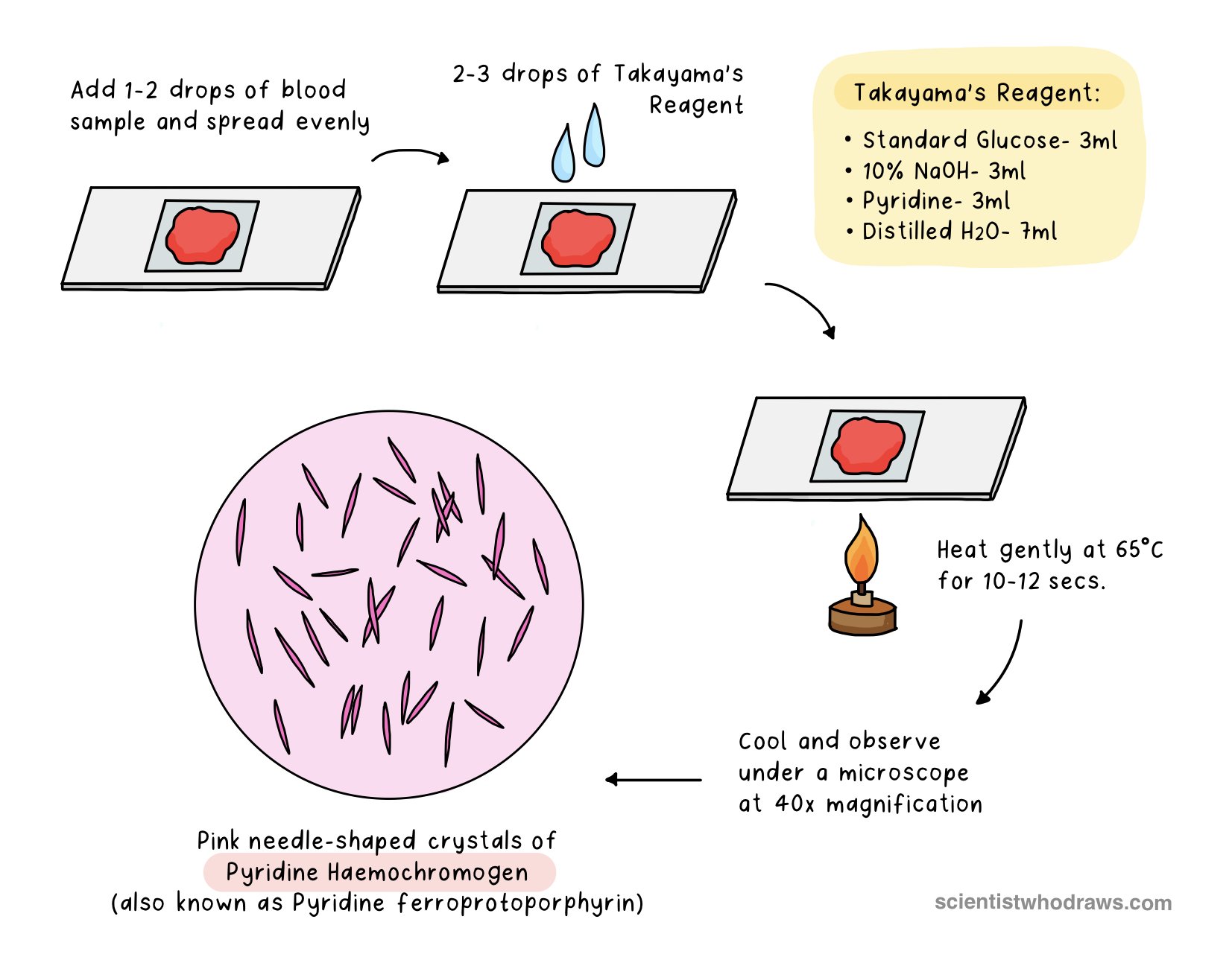
Figure: Takayama Blood Test Procedure
Principle:
Heat causes the red blood cells to rupture, releasing haemoglobin. During the process, haemoglobin gets denatured and the ferrous form of iron (Fe2+) is converted to ferric form (Fe3+) in the presence of NaOH. This oxidized haem is known as alkaline haematin. Haematin combines with pyridine to form insoluble pink needle-shaped crystals of pyridine haemochromogen (also known as pyridine ferroprotoporphyrin). In the process, saturated glucose solution functions as a reducing agent, which lowers the solubility of haemochromogen and promotes the formation of numerous crystals.
Procedure:
Put a small drop of blood on a glass slide and spread it evenly.
Add 2-3 drops of Takayama’s reagent and cover the slide with a coverslip.
Heat the slide gently at 65°C for 10-12 secs.
Keep the slide to cool at room temperature for 2-3 mins.
Observe under the microscope at 40X magnification.
Observation: Pink needle-shaped crystals of pyridine haemochromogen confirm the presence of haemoglobin in the sample.
B. TEICHMANN TEST
This test was first introduced by a Polish anatomist Ludwig Karl Teichmann in 1853. It is based on the formation of distinctive rhombus-shaped crystals of haemin when a blood sample is treated with Teichmann’s reagent.

Figure: Teichmann Blood Test Procedure
Preparation of Teichmann’s Reagent:
Dissolve 0.1 mg each of Potassium chloride (KCl), Potassium bromide (KBr), and Potassium iodide (KI) in 100ml glacial acetic acid (CH3COOH).
Principle:
KCl, KBr, and KI act as sources of chloride, bromide, and iodide ions, respectively. The chloride ions react with the iron present in hemoglobin, forming ferric chloride (FeCl3) and hydrochloric acid (HCl). The ferric chloride then reacts with hematin, a breakdown product of hemoglobin, producing a green-colored complex known as hemochromogen. Subsequently, hemochromogen reacts with HCl, resulting in the formation of a brown-colored precipitate of haemin (ferroprotoporphyrin chloride), which appears as brown rhombus-shaped crystals under the microscope. The bromide and iodide ions in the reagent also react in a similar manner with hematin, leading to the formation of a brownish precipitate of haemin.
The acetic acid in the reagent acts as a solvent to dissolve the hemoglobin and helps to stabilize the reagent and prevent the potassium salts from reacting with each other prematurely.
Procedure:
Put a small drop of blood on a glass slide and spread it evenly.
Add 2-3 drops of Teichmann’s reagent and cover the slide with a coverslip.
Heat the slide gently at 65°C for 10-12 secs.
Keep the slide to cool at room temperature for 2-3 mins.
Observe under the microscope at 40X magnification.
Observation: Brown rhombus-shaped crystals of ferroprotoporphyrin chloride (haemin) confirm the presence of haemoglobin in the sample.